Connect with us

Health

Low-cost cancer treatment with a device the size of a microwave

Published

on

Low-cost cancer treatment with a device the size of a microwave

Low-cost cancer treatment with a device the size of a microwave. A Belgian biotech company is testing a device that produces cancer drugs in hospitals, reducing waiting times and the cost of treatment.

Low-cost cancer treatment with a device the size of a microwave

In this article we’re going to read about Low-cost cancer treatment with a device the size of a microwave. When cancer treatment with a chimeric antigen receptor T cell, or CAR-T, works, it can seem miraculous. About half of leukemia and lymphoma patients, and about a third of myeloma patients, get a complete cure with a single injection of immune T cells that have been genetically modified to find and kill cancer in the blood. CAR-T treatment for acute lymphoblastic leukemia, which is the most common type of childhood cancer, has shown a cure rate of up to 90%. The first two patients treated with CAR-T in 2010 were adult men suffering from end-stage acute lymphoblastic leukemia. They were still in remission a decade after treatment.

Since 2017, the US Food and Drug Administration (FDA) has approved a total of six CAR-T therapies, all for blood cancers. Many studies have been conducted with the aim of using CAR-T therapy on solid tumors, but none are yet at the clinical trial stage. Two of these treatments, known as “Yescarta” and “Tecartus”, have earned 1.5 billion dollars for Kite Pharma in 2022 alone. Until recently, CAR-T therapies were mainly considered a last resort for patients who have tried other drugs, but CAR-T therapy can be used earlier in the treatment process and is likely to have a big impact. Last year, Yescarta was approved as a second-line treatment for large B-cell lymphoma. Despite this, drug makers are currently facing a problem.

A survey conducted in 2022 by Mayo Clinic researchers found that the average time on the waiting list for CAR-T treatment was six months, and only a quarter of patients eventually received it. Another quarter was able to enter a clinical trial for treatments that have yet to be approved. In the past few years, Bristol Myers Squibb, Kite Pharma, and Novartis have all experienced manufacturing problems with their CAR-T therapies. Johnson & Johnson (J&J) and Legend Biotech (Legend Biotech) decided in March to stop launching their CAR-T therapy, Carvycti, in the UK due to production constraints.

Unlike conventional drugs, autologous CAR-T injections are living drugs that are customized for each patient. Blood sampling of patients is usually done in a hospital or special center. Once isolated, the T cells are shipped frozen to a biomanufacturing facility where they are genetically reprogrammed to express a tumor-seeking molecule called a chimeric antigen receptor (CAR) on their surface. The modified cells are placed in an incubator for days or weeks until their numbers increase enough to create a therapeutic dose. After several stages of quality testing, the modified CAR-T cells are frozen and returned to the hospital to be injected into the patient. This process usually takes a minimum of two weeks and a maximum of eight weeks.

Current CAR-T treatments cost between $300,000 and $400,000. Travis Young, vice president of the biology department at the non-profit California Biomedical Research Institute (Calibr), said: “The reason for the high cost of treatment is that the production process must be highly controlled at every point of it.” This requires a trained technician, clean rooms, and infrastructure for transportation and freezing. The most important time is pre-release testing to ensure product sterility and potency. There are many possibilities for problems. The supply chain is still in its infancy, and it’s not just about the infrastructure, it’s about the number of people who need to be trained to do the job.

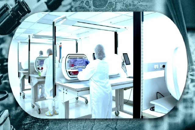
Companies are tackling these challenges in a variety of ways, aiming to reduce the complexity, time, and cost of delivering CAR-T therapies to more patients. One of the more unlikely competitors is Belgium-based Galapagos NV, which last June announced a bold plan to produce these expensive treatments faster and more cost-effectively. The program proposes developing treatments not in a centralized location, but at the point of care, using a small automated device the size of a home microwave.

Galapagos had no prior experience with CAR-T therapy and had only marketed one product in Europe, the UK, and Japan since its inception in 1999. This product was the drug “Jyseleca” for the treatment of ulcerative colitis and rheumatoid arthritis, whose sales in 2022 were reported to be equal to 95 million dollars. The drug has recently undergone a series of clinical trials, but it has one special advantage: Paul Stoffels, the new CEO of Galapagos and former chief scientific officer of Johnson & Johnson.

When Stoffels left J&J at the end of 2021, he had one of the most enviable track records in the pharmaceutical industry. This Belgian-born doctor and specialist in infectious diseases during his training was in charge of the groups that produced 25 new drugs; including two successful cancer drugs, breakthrough treatments for HIV and tuberculosis, and vaccines for Ebola and Covid-19. Although Carvycti was approved a few months after Stoffels left, it was developed under his watch. During Stoffels’ tenure, J&J’s pharmaceutical sales more than doubled from $22.5 billion in 2009 to $45.6 billion in 2020. Seven of the drugs developed under Stoffels’ supervision have been added to the “World Health Organization’s” (WHO) list of essential drugs, which means that they are considered necessary to maintain health.

Stoffels’ stint in the Galapagos gives him the opportunity to demonstrate his ability in a larger company. Immediately after taking over as CEO last April, Stoffels orchestrated a major pivot, buying two startups working on different aspects of CAR-T therapies and manufacturing them, and four months later hired 200 people working on drug programs. They were working older, fired.

Galapagos sets up manufacturing units at each of its partner hospitals, which includes training people, installing equipment, and validating the manufacturing process, Stoffels explained about the process. This is a new approach but much simpler than centralized manufacturing. In centralized manufacturing, you have to invest several hundred million dollars in a building, hire between 500 and 1,000 people, train them, and produce the drug there, but for us, the heavy lifting of this technology has already been done.

From a biological perspective, using diseased cells that have never been frozen has advantages that affect cell health. Newly generated CAR-T cells, after re-injection into the patient, show robust and consistent growth, which helps minimize a common side effect of CAR-T therapy called cytokine release syndrome, Stoffels continued. This syndrome is an aggressive reaction to immunotherapy that causes fever, nausea, and fatigue. The fact that the treatment can be done in seven days allows people with a very short life expectancy to receive this type of treatment. The first patient treated with CAR-T, who came to the hospital with acute respiratory distress syndrome and severe tumor recurrence, is still in excellent condition. This could never have been done with CAR-T the old way and in one centralized location.

Read More: Scientists discovered the secret of DNA’s X shape

Decentralization, simplification, and automation of the entire process will significantly reduce CAR-T costs, Stoffels added. The time required and the amount of work that needs to be done make CAR-T treatments expensive. If you put four or five systems in a hospital room, you can treat 200 patients a year using only a small staff.

Galapagos will not be immune to shortages of chemical reagents and other raw materials that affect other companies. “All the challenges are compounded by not having trained technicians to do the work,” Travis Young said. Technicians don’t need a lot of training because the systems require a lot less manipulation, but whenever you distribute these systems across hospital centers, you lose some control over them all.

After all, Stoffels has made the impossible possible before, and he’s done it many times, and he doesn’t seem to be giving up. He added: “I have worked all my life trying to get access to medicines.” This work is also such a mission. New science allows us to do new, difficult, and different things, and if you don’t start, you will never reach the goal.


Health

Transforming invasive cancer cells into healthy cells!

Published

on

By

Transforming invasive cancer cells into healthy cells!

Transforming invasive cancer cells into healthy cells! A specific form of aggressive childhood cancer that forms in muscle tissue may be a new cancer treatment option.

Transforming invasive cancer cells into healthy cells!

Scientists have successfully stimulated rhabdomyosarcoma cells to transform into normal, healthy muscle cells. It’s a breakthrough that could see the development of new treatments for this brutal disease and could lead to similar advances for other types of human cancer.

Rhabdomyosarcoma (RMS) is a highly aggressive type of cancer that arises from mesenchymal cells that have failed to fully differentiate into skeletal muscle myocytes. The tumor cells are identified as rhabdomyoblasts.

“These cells literally turn into muscle, and the tumor loses all of its cancerous characteristics,” says Christopher Wacock, a molecular biologist at Cold Spring Harbor Laboratory. They change from a cell that just wants to use itself more to a cell that is dedicated to contraction, and because all of its energy and resources are now devoted to contraction, it can no longer go back to proliferative and cancerous.

Read More: The genetic signal controlling the blood-brain barrier was discovered

Cancer occurs when cells in different parts of the body mutate. Rhabdomyosarcoma is a type of cancer that is mostly seen in children and teenagers. It usually starts in skeletal muscle when the cells in it mutate and begin to multiply and take over the body.

Rhabdomyosarcoma cancer is aggressive and often fatal, and the survival rate for the moderate-risk group is between 50 and 70%.

One of the promising treatment options for this disease is called “differentiation therapy”. This treatment emerged when scientists realized that leukemia cells are not fully mature and resemble undifferentiated stem cells that have not yet fully transformed into a specific cell type. Differentiation therapy forces those cells to continue growing and differentiate into specific adult cell types.

In a previous study, Wacock and colleagues effectively reversed the mutation in cancer cells that appear in Ewing’s sarcoma.

Ewing’s sarcoma is another cancer that usually appears in the bones in childhood, and is a rare, small, round, blue-colored tumor that occurs in the bones or soft tissue. This tumor can appear in any bone, but it mostly grows in the hip, thigh, arm, shoulder, and ribs, and the age of its prevalence is in adolescence or young adulthood. Ewing is slightly more common in boys than in girls.

The researchers wanted to see if they could replicate their success with rhabdomyosarcoma. At first, they thought that the realization and use of “differentiation therapy” was decades away.

The researchers used a genetic screening technique to narrow down the genes that might force rhabdomyosarcoma genes to continue growing in muscle cells. They found the answer in a protein called nuclear transcription factor-Y (NF-Y).

Rhabdomyosarcoma cells produce a protein called PAX3-FOXO1, which stimulates the proliferation of the cancer and the cancer depends on it.

The researchers found that removing the NF-Y protein inactivated the PAX3-FOXO1 protein, which in turn forced the cells to continue growing and differentiating into mature muscle cells without any signs of cancer activity.

According to the team, this is a key step in the development of a differentiation therapy for rhabdomyosarcoma and could accelerate the realization and expected timing of such therapies.

The researchers say that the positive impact of their technique, which has now been shown on two different types of sarcoma, can be applied to other sarcomas and cancer types as well, as it provides scientists with the tools needed to find out how cancer cells differentiate.

“Every successful treatment has its own story, and research like this is like fertile soil from which new drugs and treatments are born,” says Vakok. 

This research has been published in the journal of the National Academy of Sciences.

Continue Reading

Health

Inventing a lozenge to relieve tooth sensitivity

Published

on

By

Inventing a lozenge to relieve tooth sensitivity

Inventing a lozenge to relieve tooth sensitivity. Scientists have developed a way to restore the lost minerals in teeth that cause them to be sensitive.

Inventing a lozenge to relieve tooth sensitivity

What’s worse than not being able to eat delicious treats like ice cream because we don’t want to endure the pain of the cold hitting our sensitive teeth again?

This problem will soon be a thing of the past as researchers have developed a new method of restoring lost tooth minerals that offers a long-term solution to this problem.

Tooth sensitivity, also called dentin hypersensitivity, occurs when the inner dentin layer of the tooth and the tubules within it are exposed, often due to the loss of the tooth’s protective enamel in a process called demineralization. ) they say.

Read More: Low-cost cancer treatment with a device the size of a microwave

With the opening of the softer space of the tooth, its nerves, and blood vessels are prone to react to heat, cold, touch, pressure, or acidic foods, which causes pain.

Tooth enamel can be worn away by wear, decay, or grinding and cannot be repaired by natural processes as it is the only non-living tissue in our body.

In recent years, the increase in peroxide-based teeth whitening products has exacerbated tooth enamel wear, and currently, the only way to treat dentin hypersensitivity is to prevent it and treat its symptoms.

Now researchers at the University of Washington have developed a new treatment that can restore lost tooth minerals and provide a permanent solution to the problem of dentin hypersensitivity.

“We [dentists] see patients with sensitive teeth, but we can’t really help them,” says study co-author Sammy Duggan. We all have these restorative options on the market, but they are all temporary because they focus on treating the symptoms and not addressing the root cause.

The goal of researchers is to create a biosimilar, something that closely resembles or mimics the natural biochemical processes that occur in the body. So they focused on a peptide—a short chain of amino acids—that is key to the biological development of human teeth. This peptide, called sADP5, binds to calcium and phosphate ions, the main minerals found in teeth, and uses them to build new mineral microlayers.

In preclinical experiments, the researchers created a small tablet with a core of calcium and phosphate coated in the flavoring agent sADP5, which they tested on dentin discs extracted from human teeth.

Each of the discs had ivory tubes. After three rounds of peptide-guided treatment, the researchers managed to form a new mineral layer on the exposed dentin that stretched into the dentinal tubules and blocked them.

“Our technology recreates the same minerals found in teeth, including enamel, cementum, and dentin, that were previously dissolved through demineralization,” said Deniz Yusisoy, lead author of the study.

He added: the newly formed mineral microlayers close the channels of communication with the nerves of the tooth, and after that excessive sensitivity does not cause a problem anymore.

By measuring the hardness of the newly formed mineralized layer, the researchers found that it was significantly harder than non-mineralized, natural human dentin. Also, by testing it using the thermal wear method, the mineral layer was not separated from the tooth.

Both of these findings show that this method can provide resistance to long-term mechanical and thermal stresses that teeth face in the natural environment of the mouth. In addition to tablets, researchers have included their peptide-based formula in mouthwashes, tooth gels, teeth whiteners, and toothpaste.

Hanson Fong, one of the authors of the study, says: “There are many methods of design and delivery. The most important thing is the peptide, which is a key ingredient in our formula and works.

Further research is needed to investigate the permeability and chemical stability of the mineral layer to achieve an effective and easy-to-use treatment for dentin hypersensitivity, including the implementation of the peptide-guided approach in vitro.

The study was published in the journal ACS Biomaterials Science & Engineering.

Continue Reading

Health

The genetic signal controlling the blood-brain barrier was discovered

Published

on

By

The genetic signal controlling the blood-brain barrier was discovered

The genetic signal controlling the blood-brain barrier was discovered. In a new study, researchers have succeeded in identifying a genetic signal that controls the blood-brain barrier.

The genetic signal controlling the blood-brain barrier was discovered

New research in mice and zebrafish has discovered the genetic signal needed to form and maintain the blood-brain barrier.

The discovery could allow scientists to control the permeability of the blood-brain barrier and provide a more effective way to deliver drugs to the brain to treat stroke, neurological and psychiatric diseases, and cancer.

The blood-brain barrier (BBB) is a highly interconnected system of specialized cells that form a layered, semipermeable membrane that serves a dual purpose: protecting against toxins or pathogens entering the brain from the bloodstream while allowing passage through itself. gives vital nutrients.

Read More: The relationship between schizophrenia and mother’s vitamin D!

The blood-brain barrier is the separating area between the extracellular fluid of the brain in the central nervous system and the circulating blood flow in the body so that if colored substances are injected into the blood, it can be seen that there is no trace of this substance inside the brain. This curtain or barrier is made up of special capillaries, which, unlike the normal structure of capillaries, do not have the usual pores, and the intercellular connection in them is tight, and as a result, many molecules and micromolecules, as well as bacteria, are able to pass through them (through Diffusion) and reaching the cerebrospinal fluid is not in the brain. Conversely, the endothelial surface of these capillaries is covered with special proteins that allow glucose to enter the brain as nutrition. Also, gas exchange (oxygen-carbon dioxide) between circulating blood and the brain can be done without any problem through this barrier.

But the protective function of the blood-brain barrier can prevent effective drugs from being delivered to the brain to treat cancer, stroke, or neurological diseases such as Parkinson’s or Alzheimer’s.

Over the years, various methods have been developed to increase the permeability or leakiness of the blood-blood barrier to enable the delivery of drug therapies, including the use of magnetic nanoparticles, ultrasound, and engineered fat cells.

Now, a new study by Harvard Medical School researchers has identified a gene that produces a signal necessary for the development and maintenance of the blood-brain barrier and may provide a way to control its permeability.

Researchers have long known that the permeability of the blood-brain barrier is controlled by surrounding cells, but the genes in those cells remain unknown. Of course, when the researchers in the current study began to investigate the blood-brain barrier in zebrafish, the answers to these questions became clear.

In previous studies on transparent zebrafish, researchers discovered a gene called mfsd2aa that, when mutated, caused the blood-brain barrier to leak throughout the brain. But in some zebrafish, this barrier was permeable only in the forebrain and midbrain, not in the hindbrain.

“This observation led me to find a gene that makes the blood-brain barrier more permeable,” says Natasha O’Brown, lead author of the study.

In the present study, the researchers conducted additional experiments on zebrafish and mice. They found that region-specific breakdown of the blood-brain barrier is associated with mutations in the spock1 gene, which is expressed in nerve cells throughout the retina, brain, and spinal cord, but not in cells that form the blood-brain barrier.

They observed that spock1 mutant animals had more vesicles in their endothelial cells, which are key components of the blood-brain barrier. Vesicles are bubble-like membranes that store and transport cellular products and can transport large molecules across the blood-brain barrier. They also have a smaller basement membrane, which is a network of proteins found between endothelial cells and pericytes, cells that are important for forming blood vessels and maintaining the blood-brain barrier.

RNA analysis showed that spock1 alters gene expression in endothelial cells and pericytes in the blood-brain barrier, but not in other brain cell types.

When the human Spock1 protein was injected into the zebrafish brain, the endothelial cells and pericytes were repaired at the molecular level and restored about 50% of the blood-brain barrier function.

Based on this discovery, the researchers concluded that the Spock1 protein produced by neurons begins to form the blood-brain barrier during embryonic development and helps maintain it during adulthood.

“Spock1 is a potent secreted neurosignal that can promote and induce barrier properties in these blood vessels,” says O’Brown. Without it, you don’t have a functional blood-brain barrier.

The researchers say their study provides a more complete picture of the permeability of the blood-brain barrier and opens the door to the development of therapies that target spock1, potentially improving the treatment of neurological disorders such as Parkinson’s and Alzheimer’s and psychiatric disorders.

This is not the first neural signal that scientists have found, but it is the first signal from neurons that appears to specifically regulate inhibitory properties, Oberon says. I think this discovery gives us a powerful tool to try and change.

The researchers continue to look at how different pericytes are affected by spoc1 signaling. They would like to see if administering spock1 can counteract the effects of stroke on the blood-brain barrier.

This study was published in the journal Developmental Cell.

Continue Reading

Popular