
Health
The relationship between schizophrenia and mother’s vitamin D!


Why do people listen to sad songs?
There is a paradox in sad music: we don’t enjoy sadness in real life, but we enjoy art that makes us feel that way. Countless researchers since Aristotle tried to solve this contradiction. Perhaps through music, we experience a kind of catharsis of negative emotions. Catharsis here means refinement and cultivation of the soul. Maybe there is an evolutionary advantage in this feeling of sadness, or maybe we want to value our suffering. Maybe our body produces hormones in response to anxiety disorder, music that leads to a sense of comfort.
According to the New York Times, Dr. Nob, an experimental philosopher and psychologist at Yale University, in a new study published in the Aesthetic Education Journal, raised the question, “What is the purpose of sad music?” He tried to solve the contradiction of this kind of music. Over the years, he came to the conclusion that people often have two perceptions of the same thing. For example, they can consider people as artists if they have a set of characteristics such as an innate talent for working with a brush; But if they don’t have abstract values such as creativity, curiosity or interest and just recreate old masterpieces for profit, we can say they are not artists. According to Dr. Nob and his former student Tara Venkatsan, a cognitive scientist, perhaps sad music also has a dual nature.

The aforementioned research shows that our emotional response to music is multidimensional; You don’t necessarily feel happy when you listen to a beautiful song, and you don’t necessarily feel sad when you listen to a sad song. According to a 2016 study, the emotional response of 363 listeners to sad songs was divided into three categories: sadness and strong negative emotions such as anger, panic, and despair, nostalgia, quiet sadness and self-compassion, and finally sweet sadness is pleasant pain. It comes from consolation and understanding. Many respondents reported a combination of all three. The researchers called this research “Fifty Blue Spectrums”.
Given the layers of emotion and the ambiguity of language, it’s no wonder that sad music creates a paradox; But it is not clear why it induces a sense of pleasure or meaning. Some psychologists have investigated how certain aspects of music, such as position, pitch, rhythm, and resonance, are related to listeners’ emotions. According to research, certain forms of songs have an almost universal function: for example, among different countries and cultures, lullabies have similar acoustic characteristics that make children and adults feel safe. Thomas Irola, a musicologist at the University of Durham in England and researcher of the “Fifty Spectrum” study, says:
Throughout life, we learn to make connections between our feelings and what we hear. We recognize emotional expression in speech, and often these cues are used in a similar way in music.
Other researchers, such as Patrick Joslin, a music psychologist from Uppsala University in Sweden, believe that such findings reveal the value of sad music. Sad music, he writes in an essay, asks why “the second movement of Beethoven’s Eroica symphony evokes a sense of sadness?” It leads to the question, “Why does a slow step lead to a feeling of sadness?”
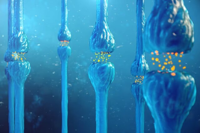
According to the findings of Joslin and his colleagues, there are cognitive mechanisms through which feelings of sadness are induced in listeners. These mechanisms include unconscious reactions in the brain stem, synchronization of the rhythm with the internal rhythm such as the heartbeat, conditional reactions to certain sounds, the arousal of memories, emotional contagion, and reflexive measurement of music. Perhaps because sadness is such a strong emotion, it can evoke an empathetic and positive response. In fact, understanding other people’s grief provokes a social response.
Why do people listen to sad songs?
The purpose of listening to sad music is not necessarily to convey sadness; Rather, it is creating a sense of connection.
Dr. Nob, along with Dr. Venkatsan and George Newman, a psychologist at the Rotman School of Management, designed a two-stage experiment to test the hypothesis. In the first part of the experiment, they gave one of four song descriptions to more than 400 participants. In the description of the first song, it was written: “transmitting complex and deep emotions, but technically full of errors.” The second track was described as: “music without technical errors that do not convey complex and deep emotions.” The third song was described as “highly emotional and technically flawless” and the fourth song was described as “technically flawed and non-emotional”.

Subjects were asked to indicate on a seven-point scale whether their song conveyed the intent of the music or not. Their goal was to show how important it is for music to express emotion and generally happiness, sadness, hate, or any other emotion on an intuitive level. Overall, subjects reported that deeply emotional but technically flawed songs best reflected the nature of music. In other words, the emotional expression had a more prominent value than the technical aspect.
In the second part of the experiment, which included 450 new subjects, the researchers gave each participant 72 descriptions of emotional songs that convey feelings such as “humiliation,” “narcissism,” “inspiration,” and “lust.” For comparison, they gave participants phrases that convey conversational interaction in expressing people’s feelings. For example, one of the phrases was: “An acquaintance is talking to you about the past week and his feeling of passion”. In general, the emotions that the subjects receive are strongly rooted in the “purpose of the music” and are similar to the emotions that make people feel close to each other in conversation: emotions such as love, joy, loneliness, sadness, ecstasy, and relaxation.
Mario Etti Picker, a philosopher at Lowell University of Chicago, finds the results of this study interesting. After reviewing the data, he came up with a relatively simple idea: “Perhaps the reason we listen to music is not just an emotional response, but we do it to understand the connection with others; Because, according to the reports of many subjects, sad music is not necessarily enjoyable despite its artistic dimensions. In other words, according to the paradox of sad music, our love of music is not the result of direct praise of sadness; Rather, it is the result of valuing communication with others.”
Dr. Irola also concluded in his research that empathic people are likely to be moved by unfamiliar sad music. They tend to engage in this kind of imaginary grief. These people also show significant hormonal changes in response to sad music. But sad music, like an onion, has many layers, and this explanation can give rise to other questions. For example, who should we communicate with? Artist or with our own past? Or even with an imaginary person?


The biography of Pavel Durov

Is Telegram really safe?

Samsung Galaxy A55 vs Galaxy S23 FE

The secret of the cleanest air on earth has been discovered

Why do people listen to sad songs?

Android 15 features: Everything you need to know 2024 update

One UI 6.1 user interface review; The most complete Android skin

Review of Xiaomi 14 Ultra, price and specifications

What would happen if gravity stopped?

Review of Motorola Edge 40 ; Pure Android in a lovely body
-
 Technology9 months ago
Who has checked our Whatsapp profile viewed my Whatsapp August 2023
-
Technology10 months ago
How to use ChatGPT on Android and iOS
-
Technology9 months ago
Second WhatsApp , how to install and download dual WhatsApp August 2023
-
Technology10 months ago
The best Android tablets 2023, buying guide
-
Humans1 year ago
Cell Rover analyzes the inside of cells without destroying them
-
AI1 year ago
Uber replaces human drivers with robots
-
Technology10 months ago
The best photography cameras 2023, buying guide and price
-
Technology10 months ago
How to prevent automatic download of applications on Samsung phones